Lower Limb Conditions

Plantar Fasciitis

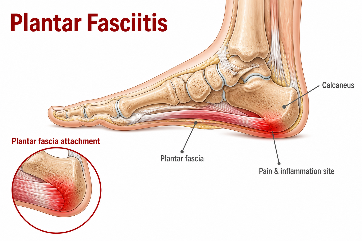
Plantar Fasciitis

Definition

Plantar Fasciitis is a common cause of heel pain involving the plantar fascia, a strong band of tissue that supports the arch of the foot. Symptoms commonly develop when the plantar fascia is exposed to more load than it can currently tolerate.

Cause

Symptoms can develop when the amount or intensity of weight-bearing activity increases beyond the plantar fascia's current capacity. Prolonged standing, changes in walking or exercise demands, and a rapid increase in activity can contribute.

Risk Factors

  • Abnormal foot-arch structure (flat feet or high arches)
  • Tight calf muscles
  • Insufficient shoe cushioning
  • Frequent changes in exercise surfaces without an adaptation period

Common Symptoms

  • Sharp stabbing pain when first stepping on the ground after getting out of bed, which eases slightly after walking
  • Deep aching pain in the heel after standing up from prolonged sitting or after prolonged standing
  • Clear tenderness at the front-inner side of the heel
  • In severe cases, pain extends across the entire foot arch and affects walking pattern

Management

Management focuses on adjusting activities that aggravate heel pain while maintaining comfortable movement. Physiotherapy may include plantar fascia and calf stretching, progressive foot and calf strengthening, and advice on activity and footwear. Persistent symptoms may require further assessment and additional treatment options.

Prevention

Choose footwear with sufficient cushioning and support, warm up thoroughly and perform dynamic stretching before exercise, increase exercise volume progressively, and use a soft mat or shift body weight regularly when working in prolonged standing.
Ankle Sprain

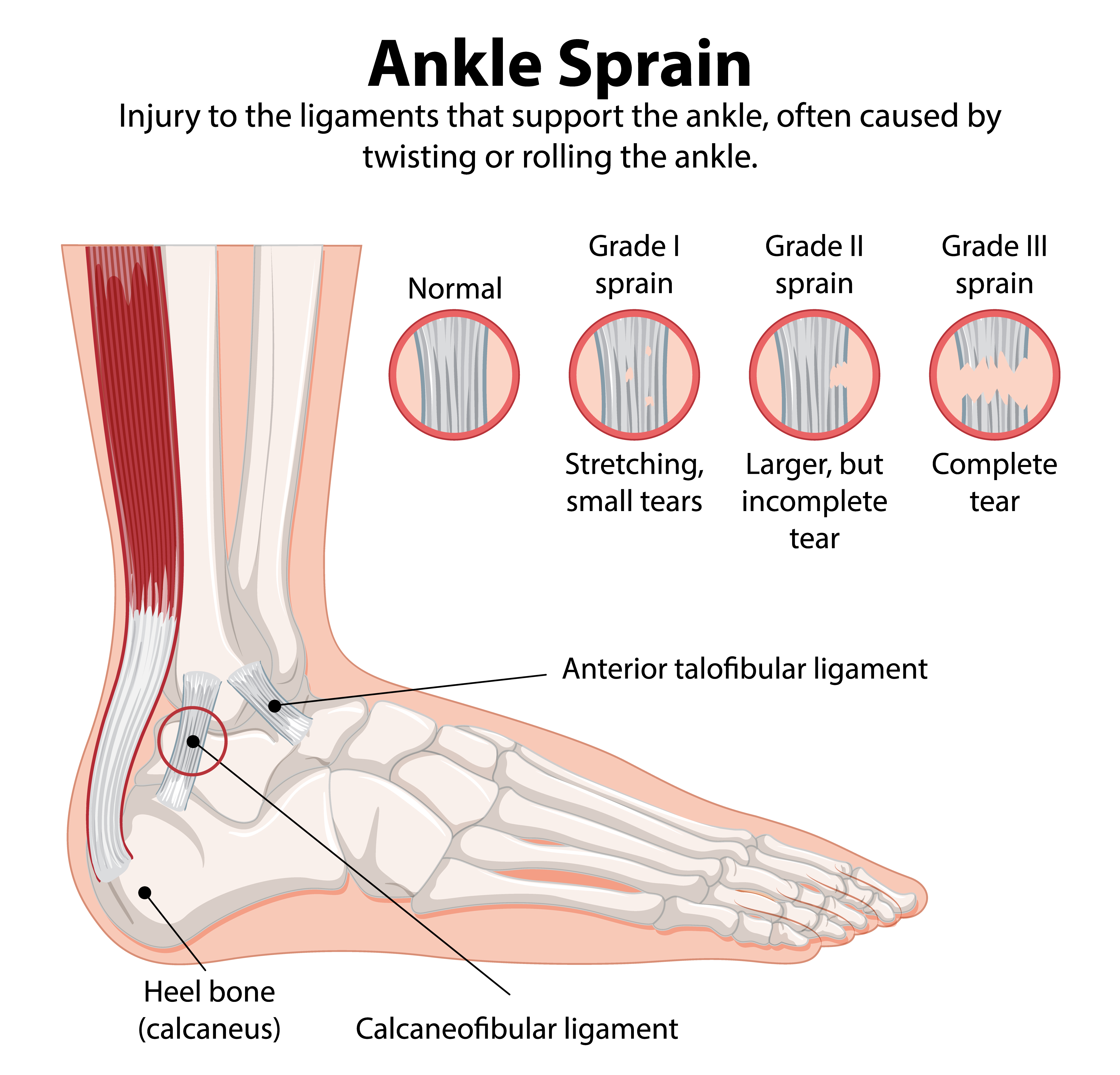
Ankle Sprain

Definition

An ankle sprain refers to injury of the surrounding ligaments caused by a sudden excessive rolling or twisting of the ankle joint. The lateral ligaments, especially the anterior talofibular ligament, are most commonly affected; severity can range from a mild strain and partial tear to a complete rupture, which may affect joint stability.

Cause

It commonly occurs when the foot suddenly inverts during landing from a jump, sudden stopping or changing direction, or stepping on uneven ground, causing the lateral ligaments to sustain excessive force. Returning to exercise too early without completing rehabilitation may reduce joint control and make recurrent sprains more likely.

Risk Factors

  • Previous ankle sprains
  • Reduced ankle mobility
  • Poor balance or proprioception
  • Lower-limb muscle weakness
  • Participation in sports involving jumping or frequent changes of direction

Common Symptoms

  • Pain, swelling, and tenderness on the outer side of the ankle; some people may develop bruising.
  • Pain worsens during weight-bearing, walking, or changing direction; severe cases may make walking difficult.
  • Reduced ankle range of motion.
  • A feeling of joint looseness or instability, or losing balance easily on uneven ground.

Management

After injury, temporarily reduce activities that significantly aggravate pain and swelling. Early rehabilitation gradually restores comfortable ankle movement and progresses ankle and calf strength, balance, proprioception, and functional control according to symptoms. If you are unable to bear weight, have marked bone tenderness, or notice deformity, seek medical assessment promptly to rule out a fracture.

Prevention

Maintain ankle and lower-limb strength, single-leg balance, and movement control. After a previous sprain, complete appropriate rehabilitation and gradually restore higher-demand activities to reduce the risk of recurrence.

Hip Osteoarthritis

Hip Osteoarthritis

Definition

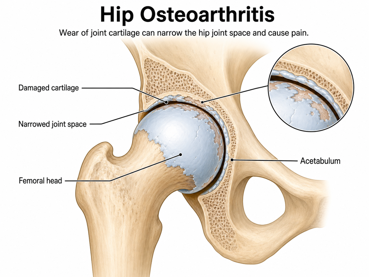
Hip osteoarthritis involves gradual degeneration of the cartilage and surrounding structures of the acetabulum and femoral head, making joint movement less smooth and potentially causing pain, stiffness, and reduced daily function. The degree of degeneration on imaging does not always fully correspond with pain or functional limitation.

Cause

Age-related degeneration, previous trauma, hip developmental abnormalities, or long-term changes in joint loading may progressively alter cartilage and other joint structures.

Risk Factors

  • Increasing age
  • Higher body weight
  • Previous hip injury or surgery
  • Joint developmental problems
  • Activities involving repeated high loading

Common Symptoms

  • Pain in the groin, front, or outer side of the hip
  • Pain that worsens with walking, stair climbing, or prolonged standing
  • Joint stiffness on waking or after prolonged sitting
  • Restricted movement when putting on shoes, getting in or out of a car, or squatting

Management

Physiotherapy may include hip mobility, gluteal and thigh strengthening, gait, and balance training according to individual needs, together with activity adjustment and weight management. When pain continues to affect daily life, a doctor should assess medication, injections, or surgery; treatment progression should be adjusted according to pain, function, and individual goals.

Prevention

Maintaining an appropriate body weight, continuing hip strengthening and mobility exercises, and progressively adjusting high-impact exercise volume can help maintain joint function.

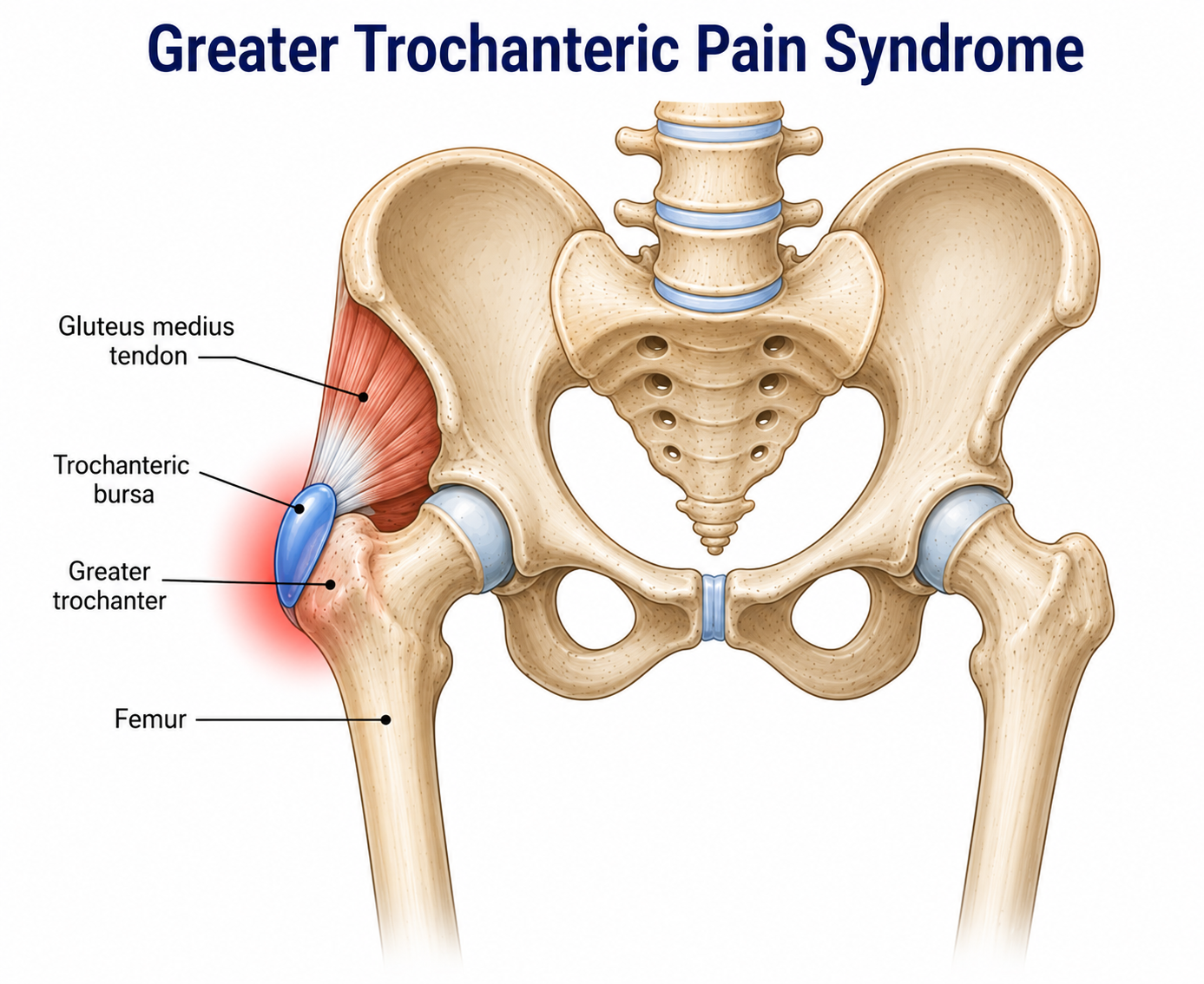
Greater Trochanteric Pain Syndrome

Greater Trochanteric Pain Syndrome

Definition

Greater trochanteric pain syndrome is pain on the outer side of the hip, often related to compression or excessive loading of the gluteus medius and minimus tendons, and sometimes involving bursal irritation. This pain is not necessarily caused by the bursa alone and is often related to loading of the gluteal tendons.

Cause

Symptoms can develop when the gluteal tendons are exposed to compression or loading beyond their current capacity. Changes in activity levels, prolonged compressive positions, and reduced hip strength or control can contribute.

Risk Factors

  • Female sex
  • Middle or older age
  • Gluteal weakness
  • Low-back or lower-limb mechanical problems
  • Prolonged side-lying on the affected side
  • A sudden increase in walking or running volume

Common Symptoms

  • Local pain and tenderness on the outer side of the hip
  • Pain when lying on the affected side
  • Worsening pain with stair climbing, uphill walking, or single-leg standing
  • Pain that may extend down the outer thigh

Management

Early management focuses on modifying positions and activities that compress or excessively pull on the gluteal tendons, while controlling exercise load. Physiotherapy progressively trains the gluteal muscles, core, and lower-limb control to restore walking, stair climbing, and exercise capacity; progression should be adjusted according to pain and function.

Prevention

Avoid prolonged weight-bearing on one leg, leg crossing, or sleeping on the affected side, and continue strengthening the gluteal and core muscles; increase activity volume gradually.

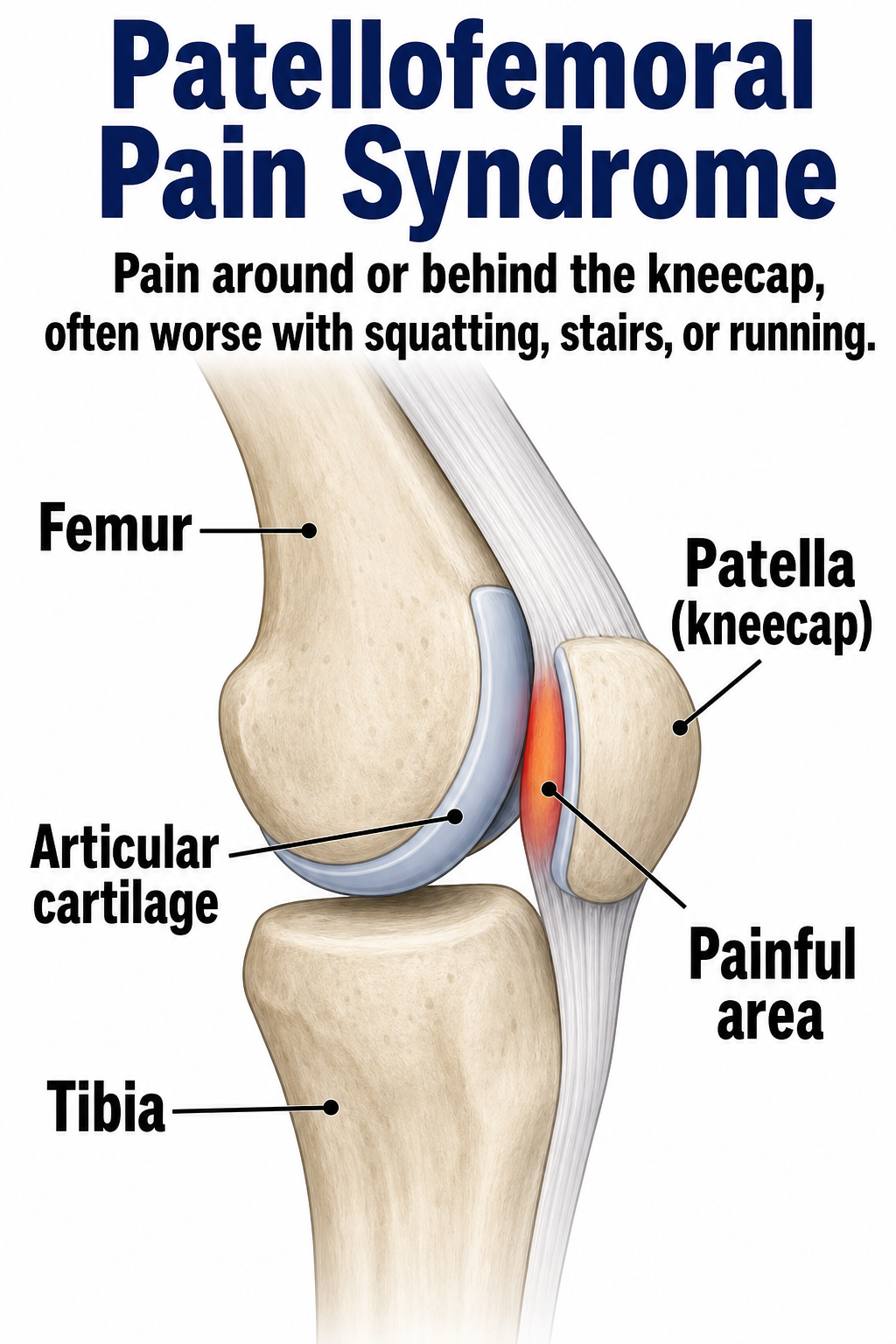
Patellofemoral Pain Syndrome

Patellofemoral Pain Syndrome

Definition

Patellofemoral pain syndrome is pain at or behind the kneecap, usually occurring when the knee is bent and weight-bearing, such as during squatting, stair climbing, running, or jumping. Pain does not necessarily indicate serious joint damage, but loading and movement should be adjusted appropriately.

Cause

Patellofemoral pain can develop when activities place greater demand on the knee than it is currently prepared for. Changes in activity level, muscle capacity, and individual movement patterns can influence symptoms.

Risk Factors

  • A sudden increase in running, jumping, or squat training volume
  • Insufficient hip or quadriceps strength
  • Poorer lower-limb movement control

Common Symptoms

  • Aching pain around or behind the kneecap
  • Pain during squatting, stair climbing, running, or jumping
  • Anterior knee discomfort when standing after prolonged sitting
  • A grinding sensation or sound during knee bending

Management

Management focuses on temporarily adjusting activities that provoke pain while gradually improving hip and knee strength and lower-limb control. Rehabilitation is progressed according to symptoms, functional needs, and the activities the person wants to return to.

Prevention

Increase running, jumping, and lower-limb training volume progressively, continue gluteal and quadriceps training, and pay attention to knee control during squats, stair climbing, and landing.

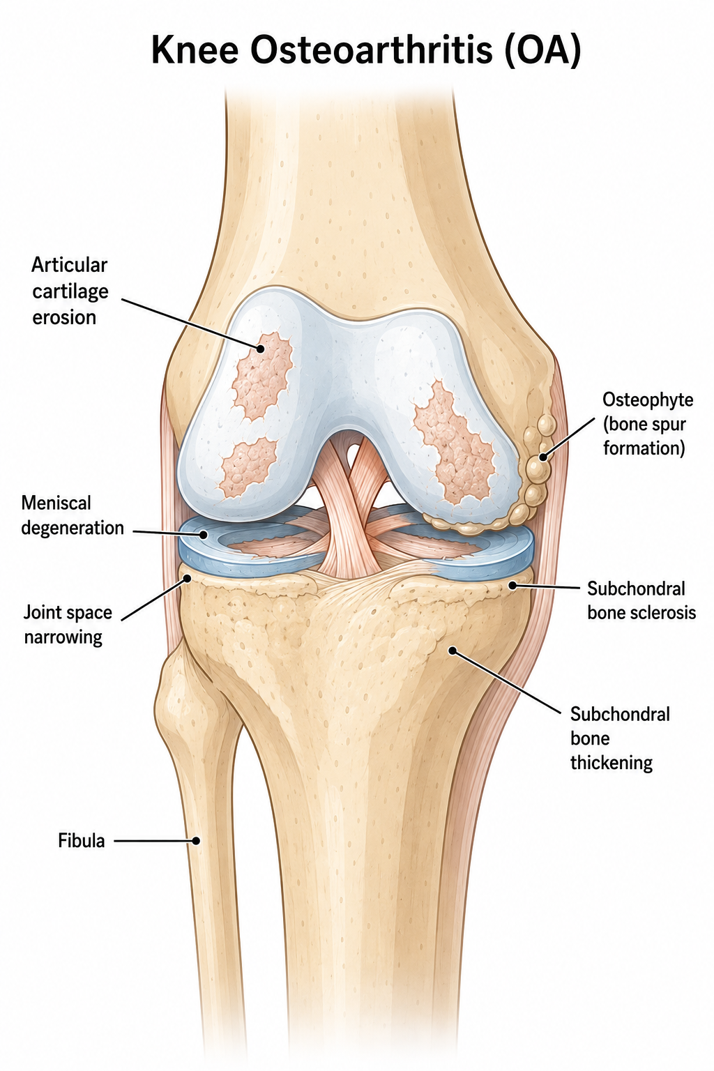
Knee Osteoarthritis

Knee Osteoarthritis

Definition

Knee osteoarthritis involves gradual degeneration of knee cartilage and surrounding structures, which may lead to pain, stiffness, reduced joint movement, and reduced walking ability. The degree of degeneration on imaging does not always fully correspond with pain or functional limitation.

Cause

Long-term accumulated joint loading and tissue degeneration influence each other; age, previous injury, or changes in joint alignment can gradually alter cartilage and other joint structures.

Risk Factors

  • Increasing age
  • Higher body weight
  • Previous knee injury or surgery
  • Repeated kneeling or high-impact activities
  • Insufficient thigh strength

Common Symptoms

  • Knee pain more noticeable after activity or weight-bearing
  • Brief stiffness on waking or after rest
  • Difficulty with stair climbing, squatting, or prolonged standing
  • Knee swelling, reduced movement, or a feeling of weakness

Management

Exercise therapy is central, including thigh and gluteal strengthening, joint mobility, walking, and balance training, together with activity adjustment and weight management. When symptoms are prominent, a doctor may assess pain medication, injections, or surgery.

Prevention

Maintain regular exercise and an appropriate body weight, progress weight-bearing activity gradually, and strengthen the thigh and gluteal muscles to help reduce knee loading and maintain function.

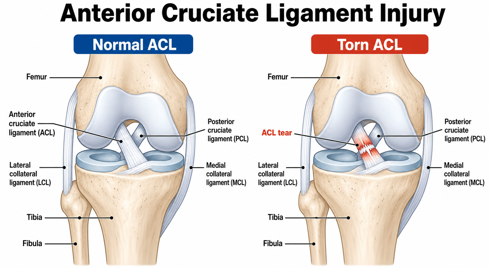
Anterior Cruciate Ligament Injury

Anterior Cruciate Ligament Injury

Definition

The anterior cruciate ligament lies within the knee joint and limits excessive forward movement and rotation of the tibia. Force beyond its capacity can cause a partial or complete tear. Some people can return to daily activities through comprehensive conservative rehabilitation; treatment plans should be individualised.

Cause

ACL injuries commonly occur when the knee is exposed to sudden twisting, rapid changes in movement, awkward landing, or direct trauma. These forces can place excessive strain on the ligament and cause a partial or complete tear.

Risk Factors

  • Sports involving sudden stopping, cutting, and jumping
  • Poor landing control
  • Lower-limb strength asymmetry
  • Insufficient core stability
  • Previous knee injury

Common Symptoms

  • Feeling or hearing a "pop" inside the knee at injury
  • Rapid joint swelling within hours
  • Knee pain and restricted bending or straightening
  • A feeling of knee instability during turning or weight-bearing

Management

The need for reconstruction depends on tear severity, joint instability, activity demands, and individual goals. Conservative or postoperative rehabilitation progressively addresses swelling, movement, strength, balance, and functional control to support a safe return to the person's required daily, work, exercise, or sporting activities.

Prevention

Train gluteal, core, and lower-limb strength, learn proper landing and cutting techniques, and include single-leg balance and movement-control training under fatigue.

Meniscal Injury

Meniscal Injury

Definition

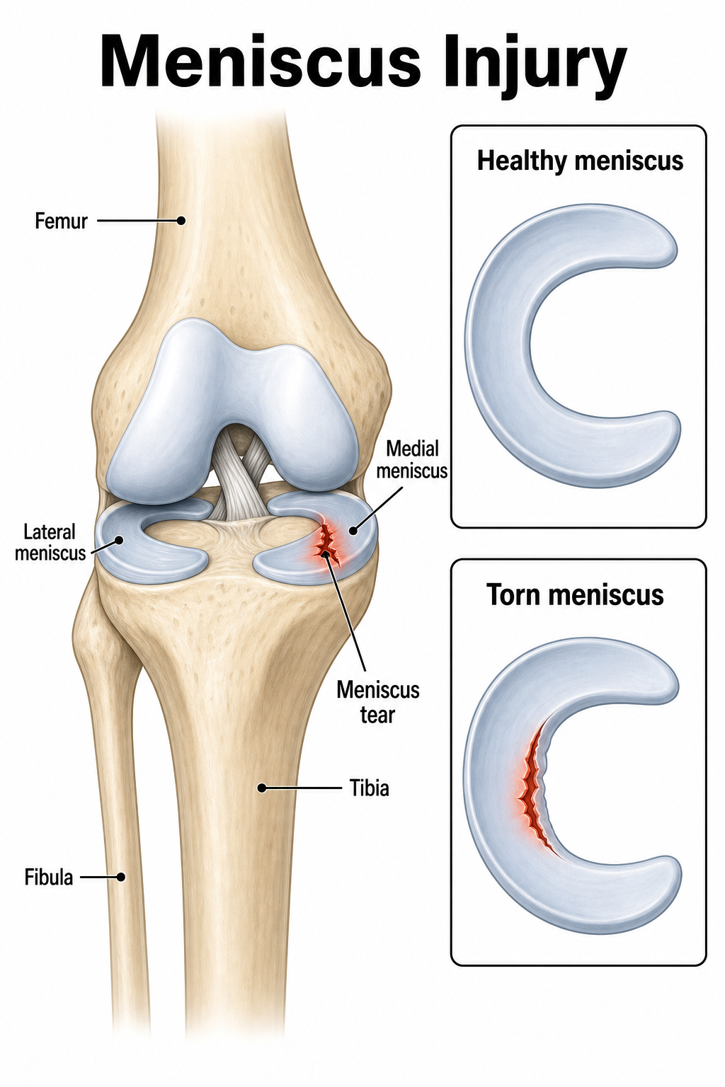
The menisci are fibrocartilage structures within the knee that help distribute load and improve stability. They may tear through acute twisting injury or long-term degeneration, and some tears interfere with smooth movement. Not every tear requires surgery.

Cause

Younger people more commonly sustain injury during twisting, squatting, or contact trauma; with increasing age, degenerative tears can occur after relatively minor movements.

Risk Factors

  • Sports involving sudden stopping and cutting
  • Previous ACL injury
  • Repeated squatting and twisting
  • Knee osteoarthritis
  • Age-related degeneration

Common Symptoms

  • Local pain on the inner or outer knee
  • Swelling or fluid in the knee after injury
  • Pain during squatting, twisting, or turning
  • Knee catching, locking, or inability to fully bend or straighten

Management

Initially, adjust activity according to symptoms while restoring knee movement and thigh strength and improving lower-limb control. Persistent locking, marked restriction, or recurrent swelling should be assessed by an orthopaedic doctor for imaging and possible surgery.

Prevention

Maintain lower-limb strength and movement control, progress demanding activities gradually, and complete appropriate rehabilitation after knee injury before returning to higher-demand activities.

Iliotibial Band Syndrome

Iliotibial Band Syndrome

Definition

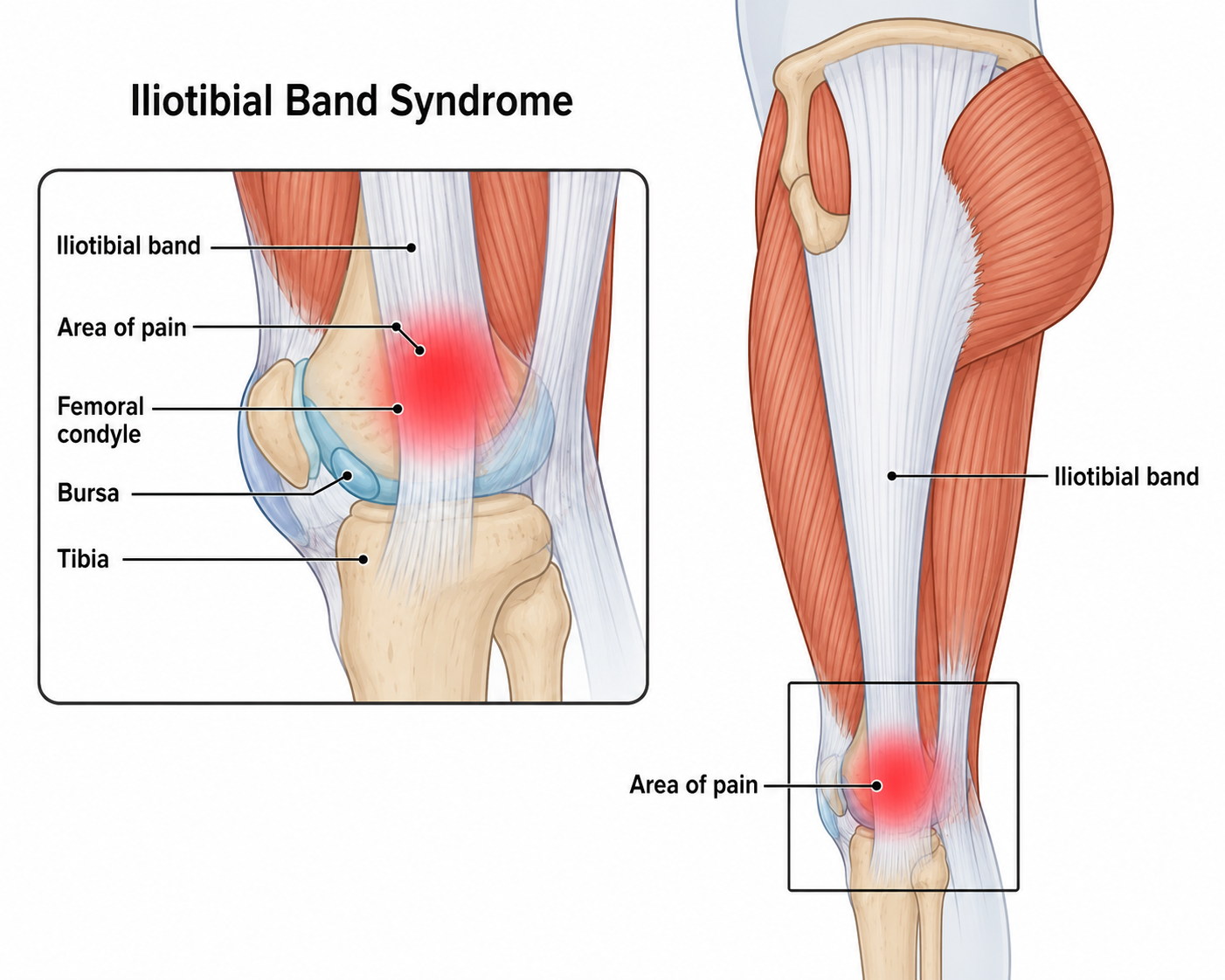
Iliotibial Band Syndrome is a load-related condition causing pain on the outer side of the knee. Symptoms commonly occur during repetitive lower-limb activity and are associated with irritation or compression of sensitive tissues around the outer knee.

Cause

Symptoms can develop when repeated lower-limb activity places greater demand on the tissues around the outer knee than they can tolerate. Changes in activity volume, terrain, and lower-limb control can contribute.

Risk Factors

  • A sudden increase in running volume
  • Downhill running
  • Sloped or uneven surfaces
  • Insufficient gluteal strength and lower-limb control
  • Unsuitable footwear or training plans

Common Symptoms

  • Local sharp or burning pain on the outer knee
  • Pain gradually appearing or worsening after running a certain distance
  • More noticeable pain during downhill walking or stair descent
  • Pain provoked by pressing on the outer knee

Management

First reduce running volume, speed, or downhill training that provokes symptoms and identify training-load problems. Physiotherapy improves hip and lower-limb strength, running control, and mobility before progressively returning to running.

Prevention

Progress running volume and intensity gradually, allow adequate recovery time, and continue training gluteal and lower-limb strength and running technique.

Tennis Leg

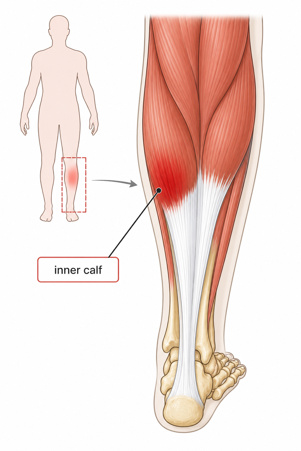
Posterior view showing the inner calf commonly affected by Tennis Leg

Definition

Tennis Leg is an acute calf injury, most commonly involving a tear near the muscle–tendon junction of the medial gastrocnemius. The soleus may also be involved, while an isolated plantaris tear is uncommon. It typically causes sudden inner-calf pain and difficulty walking or pushing through the foot.

Cause

Tennis Leg usually occurs when the calf is suddenly stretched while contracting forcefully, creating excessive strain through the medial calf. This can happen during a rapid step, push-off, sudden change in movement, or other forceful lower-limb activity.

Risk Factors

  • Previous calf strain or incomplete rehabilitation
  • Rapid increase in demanding lower-limb activity
  • Reduced calf strength or tolerance to rapid loading
  • Fatigue during repeated acceleration or direction changes

Common Symptoms

  • Sudden sharp pain in the inner or upper calf
  • A snapping or “kicked in the calf” sensation
  • Swelling or bruising that may develop after the injury
  • Pain and weakness when walking, pushing off, or performing a heel raise

Management

Physiotherapy initially protects the calf while restoring comfortable ankle movement and walking. Rehabilitation progresses from calf activation to calf raises, heavier strengthening, hopping, running, and direction-change drills. Return to sport is guided by calf strength and task performance. Increasing or unexplained swelling, redness, or warmth requires urgent assessment for a blood clot. Sudden breathlessness or chest pain requires emergency care.

Prevention

Build calf strength and tolerance to demanding lower-limb activity progressively. After injury, restore calf endurance, power, and the movements required for daily, work, or exercise activities before returning fully to higher loads.

Achilles Tendinopathy

Achilles Tendinopathy

Definition

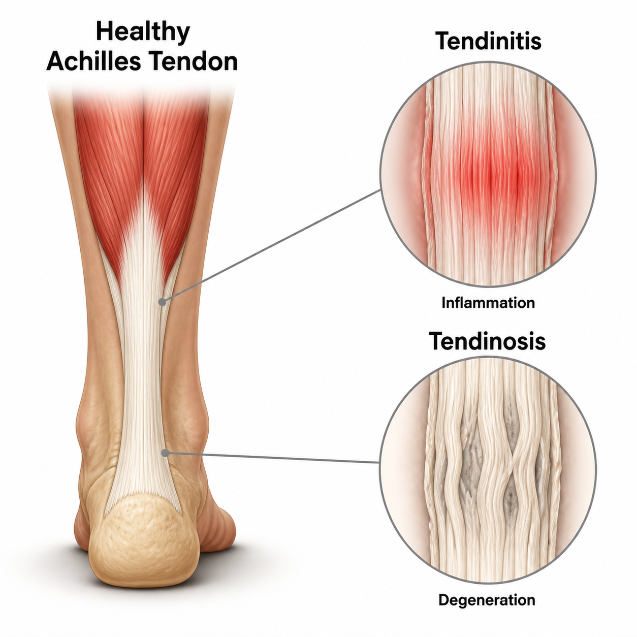
Achilles Tendinopathy is a condition affecting the Achilles tendon that commonly causes local pain, stiffness, and reduced ability to tolerate activities that load the tendon.

Cause

When running, jumping, or acceleration load exceeds the tendon's capacity to adapt, tissue response and degenerative changes can occur, often related to a rapid short-term increase in exercise volume.

Risk Factors

  • A sudden increase in running, jumping, sprinting, or uphill training
  • Reduced calf strength or mobility
  • Previous Achilles problems
  • Unsuitable footwear or insufficient recovery

Common Symptoms

  • Pain in the midportion of the Achilles tendon or behind the heel
  • Achilles stiffness on waking or after rest
  • Worsening pain with running, jumping, heel raises, or stair climbing
  • Local tendon tenderness, thickening, or swelling

Management

The central approach is to adjust running and jumping load according to pain response while progressively loading the Achilles tendon and strengthening the calf. A physiotherapist assesses ankle mobility, gait, and exercise planning to progressively restore function.

Prevention

Progress running, jumping, and speed training gradually, maintain calf strength and ankle mobility, allow sufficient recovery, and choose appropriate footwear.

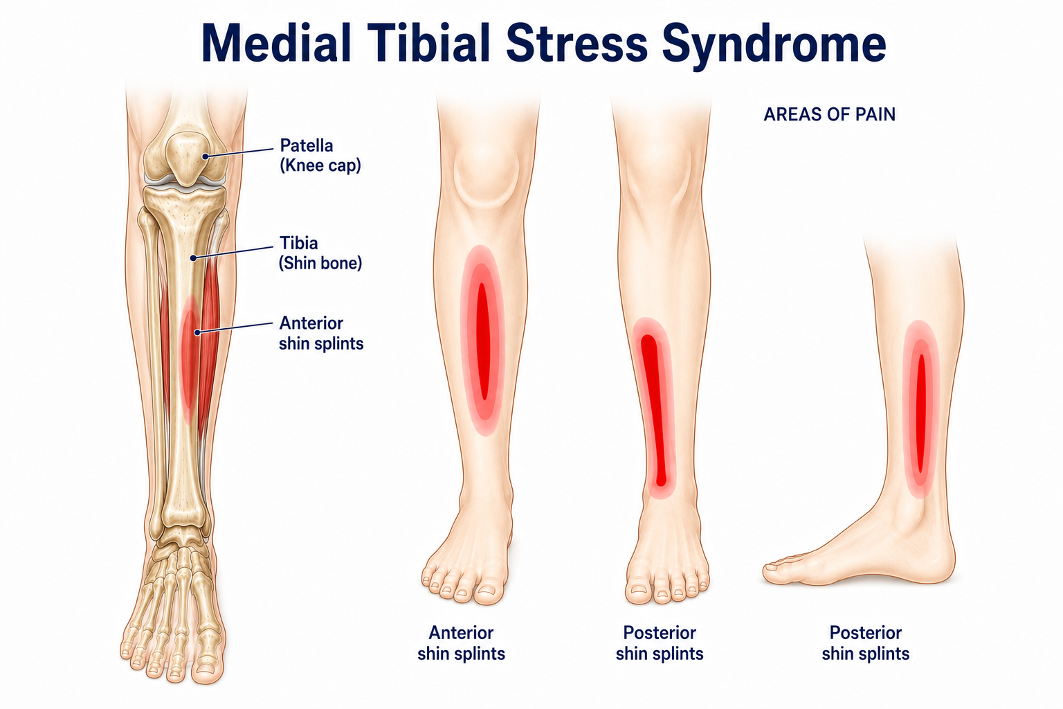
Medial Tibial Stress Syndrome

Medial Tibial Stress Syndrome

Definition

Medial Tibial Stress Syndrome is an activity-related pain condition along the inner border of the shin, associated with repeated loading of the tibia and surrounding tissues.

Cause

When running and jumping load increases faster than the body can adapt and recover, tissues around the tibia can remain irritated and progressively become painful.

Risk Factors

  • A sudden increase in running, jumping, or marching training
  • Hard surfaces
  • Excessive foot pronation
  • Tight calves
  • Insufficient lower-limb strength
  • Inadequate recovery

Common Symptoms

  • Diffuse pain or tenderness along the inner border of the tibia
  • Pain at the start of running or jumping that worsens after activity or the next day
  • Tightness or mild swelling on the inner lower leg
  • Pain during everyday walking in more severe cases

Management

Temporarily reduce running and jumping load that provokes pain and use symptom-free cross-training. Physiotherapy addresses ankle mobility, calf and foot strength, lower-limb control, and training progression; pain focused on one bone point or progressively worsening pain should be assessed for stress fracture.

Prevention

Progress running and jumping gradually, schedule recovery days, continue calf and lower-limb strengthening, and review footwear, surface, and running and jumping technique.

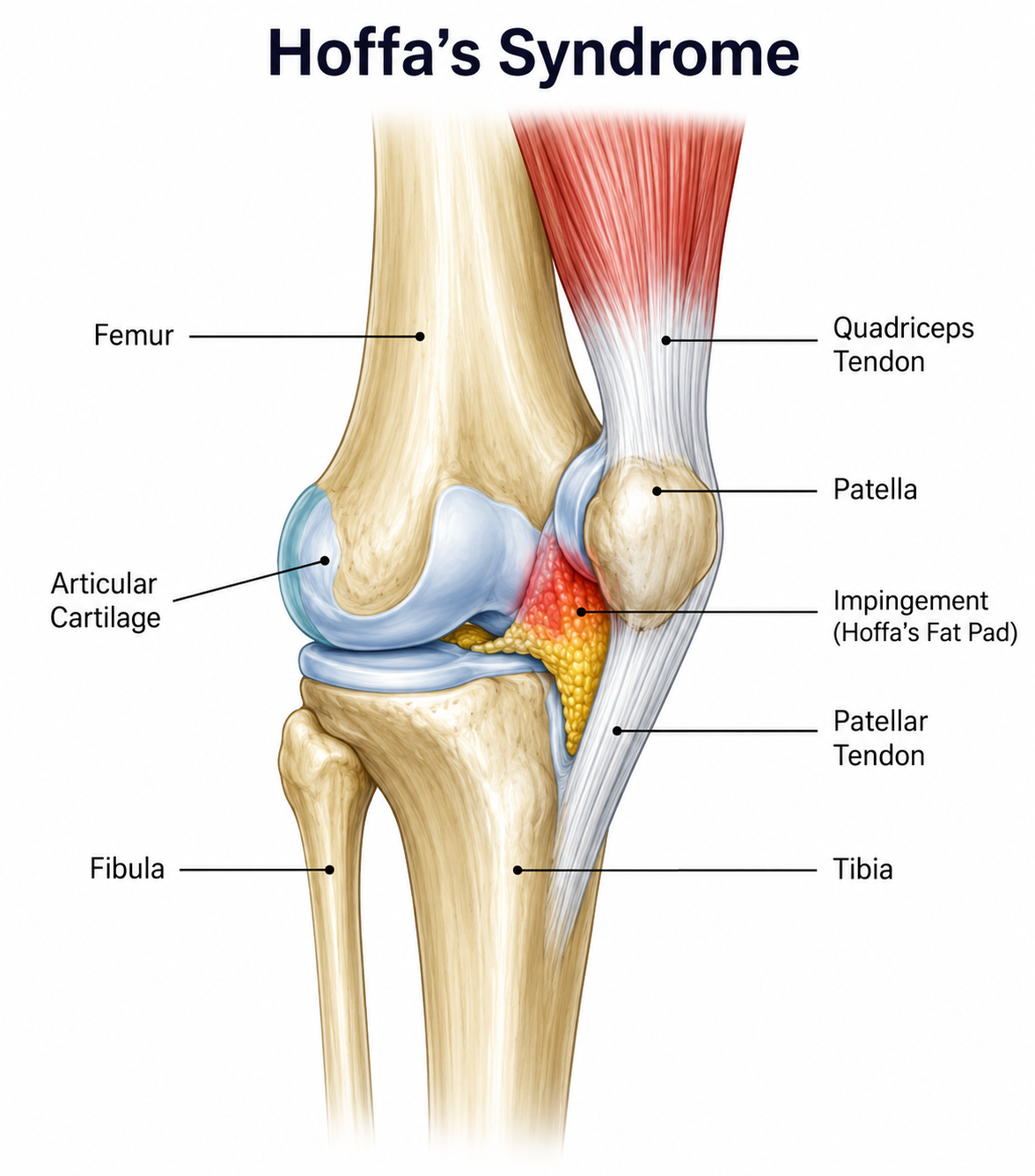
Infrapatellar Fat Pad Impingement Syndrome (Hoffa’s Syndrome)

Infrapatellar Fat Pad Impingement Syndrome (Hoffa’s Syndrome)

Definition

The infrapatellar fat pad lies below the patella and behind the patellar tendon. It is richly supplied with nerves and blood vessels and is sensitive to pain. Repeated compression, impingement, or trauma can cause swelling, inflammation, or fibrosis, resulting in anterior knee pain known as Hoffa’s syndrome.

Cause

The infrapatellar fat pad can become irritated following repeated compression, knee trauma, surgery, or repeated knee hyperextension. Changes in knee movement and loading can contribute to continued irritation and anterior knee pain.

Risk Factors

  • Knee hyperextension or a habit of locking the knees while standing
  • A sudden increase in running, jumping, kicking, or lower-limb training
  • Previous anterior-knee trauma, patellar dislocation, or knee surgery
  • Reduced hip and knee strength or movement control

Common Symptoms

  • Deep anterior knee pain below the patella or on either side of the patellar tendon
  • Worse pain with full knee extension, hyperextension, prolonged standing, or downhill walking
  • Anterior knee pain with squatting, stair climbing, running, or jumping
  • Tenderness below the patella and beside the patellar tendon, sometimes with tightness or swelling

Management

Initially, reduce activities and positions that clearly aggravate anterior knee pain, particularly repeated knee hyperextension. Physiotherapy gradually restores comfortable knee movement and develops quadriceps, hip, balance, and lower-limb control according to symptoms and functional needs. Persistent pain, knee locking, or inability to bear weight after trauma requires medical assessment.

Prevention

Avoid standing with the knees locked for long periods and progress running, jumping, squatting, and lower-limb resistance training gradually. Continue strengthening the quadriceps, gluteal, and core muscles, and keep the knee aligned over the second toe during landing, stair descent, and single-leg loading to reduce hyperextension and dynamic valgus stress.

Book
Ask